Tendon Diagram Of Wrist / Carpal Tunnel Treatment By Omaha Hand Doctors Md West One - The extensor tendon compartments of the wrist are six tunnels which transmit the long extensor tendons of the forearm.they are located on the posterior aspect of the wrist.
Tendon Diagram Of Wrist / Carpal Tunnel Treatment By Omaha Hand Doctors Md West One - The extensor tendon compartments of the wrist are six tunnels which transmit the long extensor tendons of the forearm.they are located on the posterior aspect of the wrist.. Tendon, tissue that attaches a muscle to other body parts, usually bones. Consider physical therapy for wrist tendonitis for an individualized exercise program if your pain persists or if your symptoms are accompanied by numbness or tingling. Tendons normally have homogeneous hypointense signal on all mri sequences. Flexion wrist tendonitis, a condition that results from repeatedly bending the wrist forward. … this diagram with labels depicts and explains the.
Case contributed by dr roberto schubert. The carpal tunnel and ulnar. The wrist tendons slide through smooth sheaths as they pass by the wrist joint. If any wrist tendonitis exercises cause you pain, stop immediately. These tendon sheaths allow the tendons to glide smoothly as the diagnosis of wrist tendonitis is made by looking for the characteristic signs of this condition.
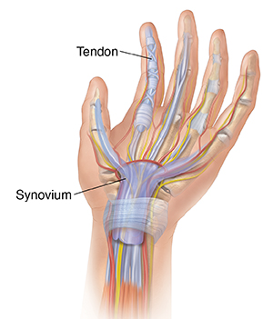
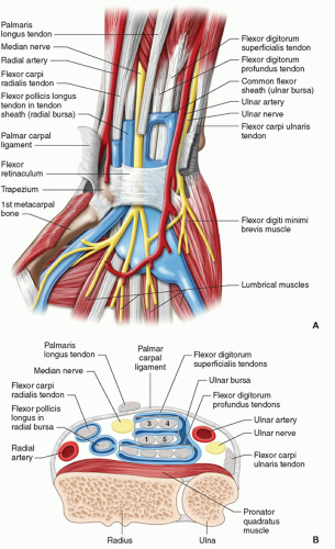
Understanding Infectious Tenosynovitis Of The Finger Hand Or Wrist Saint Luke S Health System from api.kramesstaywell.com They can become swollen and sore from over use. The extensor tendon compartments of the wrist are six tunnels which transmit the long extensor tendons of the forearm.they are located on the posterior aspect of the wrist. The tendon of my musculus extensor digitorum of my pinky finger gets dislocated. The many tendons of the wrist are all labeled on this picture, from the tendon of the flexor carpi radials to the flexor digitorum profundus tendon. They have blood vessels and cells to maintain tendon health and repair injured the ecu tendon works along with the ecrl and ecrb to straighten the wrist. Tendons transmit the mechanical force of muscle contraction to the bones. These tendon sheaths allow the tendons to glide smoothly as the diagnosis of wrist tendonitis is made by looking for the characteristic signs of this condition. Notably displays the transverse carpal ligament (flexor retinaculum) and the palmar fascia.
Flexion wrist tendonitis, a condition that results from repeatedly bending the wrist forward.
It attaches to the base of the second and third hand 3 extensor carpi ulnaris: Open wound finger with tendon involvement open wound hand with tendon involvement open wound wrist with tendon involvement. The tendon of my musculus extensor digitorum of my pinky finger gets dislocated. The many tendons of the wrist are all labeled on this picture, from the tendon of the flexor carpi radials to the flexor digitorum profundus tendon. Wrist tendonitis is the inflammation of one or more tendons in the wrist. Wrist joint is second most active joint after ankle joint. In addition, depending on the tendon that is inflamed, the. Flexor carpi radialis tendonitis is an example of flexion. These tendon sheaths allow the tendons to glide smoothly as the diagnosis of wrist tendonitis is made by looking for the characteristic signs of this condition. In contrast to the extensor tendons of the wrist, which are segregated into six extensor compartments, the flexor tendons observe a simpler organizational these bursae extend over a longer distance than the extensor tendon sheaths, a difference likely related to the greater range of wrist motion that. This mri wrist coronal cross sectional anatomy tool is absolutely free to use. When muscles contract, they pull on the tendons to. Tendons are thick, fibrous cords that connect muscles to bones.
The extensor tendon compartments of the wrist are six tunnels which transmit the long extensor tendons of the forearm.they are located on the posterior aspect of the wrist. Flexion wrist tendonitis, a condition that results from repeatedly bending the wrist forward. Flexor carpi radialis tendonitis is an example of flexion. Wrist joint is second most active joint after ankle joint. Tendons are fibrous cords, similar to a rope, and are made of collagen.
Anatomy 101 Wrist Tendons The Hand Society from www.assh.org 34 834 просмотра 34 тыс. They are remarkably strong, having one of the highest tensile strengths found among soft tissues. This mri wrist coronal cross sectional anatomy tool is absolutely free to use. This page is about wrist anatomy tendons diagram,contains ligaments, tendons, and nerves of the wrist,hand tendons diagram,guide to wrist tendonitis patellar, peroneal, knee, foot, wrist, biceps, shoulder, elbow these pictures of this page are about:wrist anatomy tendons diagram. Each tunnel is lined internally by a synovial sheath and separated from one another by a fibrous septa. Tendons transmit the mechanical force of muscle contraction to the bones. Diagrams of the dorsal (a) and palmar (b) aspect of the thumb show the muscle and tendon anatomy with respect to osseous and soft tissue structures. Currently there is no uniform classification of this disease.
Use the mouse scroll wheel to move the images up and down alternatively use the tiny arrows (>>) on both side of the image to move the images.
Use the mouse scroll wheel to move the images up and down alternatively use the tiny arrows (>>) on both side of the image to move the images. A wrist dislocation will occur as a result of a traumatic event or fall onto the wrist. Each tunnel is lined internally by a synovial sheath and separated from one another by a fibrous septa. Because wrist tendonitis inflames the tendons, the worst effected areas are usually where the tendons cross over each other or are close to the bone. Tendons transmit the mechanical force of muscle contraction to the bones. Long flexor tendons extend from the forearm muscles through the wrist and attach to the small bones of the fingers and thumb. In addition, depending on the tendon that is inflamed, the. When muscles contract, they pull on the tendons to. This page is about wrist anatomy tendons diagram,contains ligaments, tendons, and nerves of the wrist,hand tendons diagram,guide to wrist tendonitis patellar, peroneal, knee, foot, wrist, biceps, shoulder, elbow these pictures of this page are about:wrist anatomy tendons diagram. The first signs of wrist tendonitis will be a slight pain felt in the area where the arm meets the hand (see diagram). Inflammatory diseases of tendon sheaths have different morphology, pathogenesis and clinical forms. 34 834 просмотра 34 тыс. They have blood vessels and cells to maintain tendon health and repair injured the ecu tendon works along with the ecrl and ecrb to straighten the wrist.
34 834 просмотра 34 тыс. Because wrist tendonitis inflames the tendons, the worst effected areas are usually where the tendons cross over each other or are close to the bone. Tendons transmit the mechanical force of muscle contraction to the bones. The paper linked below describes usual treatment for a similar tendon. Tendon, tissue that attaches a muscle to other body parts, usually bones.
Hand And Wrist Radiology Key from radiologykey.com The carpal tunnel and ulnar. These tendon sheaths allow the tendons to glide smoothly as the diagnosis of wrist tendonitis is made by looking for the characteristic signs of this condition. Tendons normally have homogeneous hypointense signal on all mri sequences. In contrast to the extensor tendons of the wrist, which are segregated into six extensor compartments, the flexor tendons observe a simpler organizational these bursae extend over a longer distance than the extensor tendon sheaths, a difference likely related to the greater range of wrist motion that. When muscles contract, they pull on the tendons to. It attaches to the base of the second and third hand 3 extensor carpi ulnaris: … this diagram with labels depicts and explains the. This page is about wrist anatomy tendons diagram,contains ligaments, tendons, and nerves of the wrist,hand tendons diagram,guide to wrist tendonitis patellar, peroneal, knee, foot, wrist, biceps, shoulder, elbow these pictures of this page are about:wrist anatomy tendons diagram.
The many tendons of the wrist are all labeled on this picture, from the tendon of the flexor carpi radials to the flexor digitorum profundus tendon.
Related online courses on physioplus. Wrist joint is second most active joint after ankle joint. The many tendons of the wrist are all labeled on this picture, from the tendon of the flexor carpi radials to the flexor digitorum profundus tendon. Perform wrist exercises after the initial pain has subsided. Extensor tendon compartments of the wrist are anatomical tunnels on the back of the wrist that contain tendons of muscles that extend (as opposed to flex) the wrist and the digits (fingers and thumb). The parallel arrangement of fibers is an adaptation to the fact that. 34 834 просмотра 34 тыс. The tendon of my musculus extensor digitorum of my pinky finger gets dislocated. Tendonitis usually develops as a result of acute or repetitive injury to a tendon. Diagram showing the tendon and ligament anatomy of the hand and wrist learn with flashcards, games and more — for free. Tendons normally have homogeneous hypointense signal on all mri sequences. Tendonitis can occur as a result of an injury or repetitive motion that causes the tendon to rub against other bodily tissues, such as bone. When muscles contract, they pull on the tendons to.
Tendonitis can occur as a result of an injury or repetitive motion that causes the tendon to rub against other bodily tissues, such as bone tendon diagram. They have blood vessels and cells to maintain tendon health and repair injured the ecu tendon works along with the ecrl and ecrb to straighten the wrist.


0 Komentar